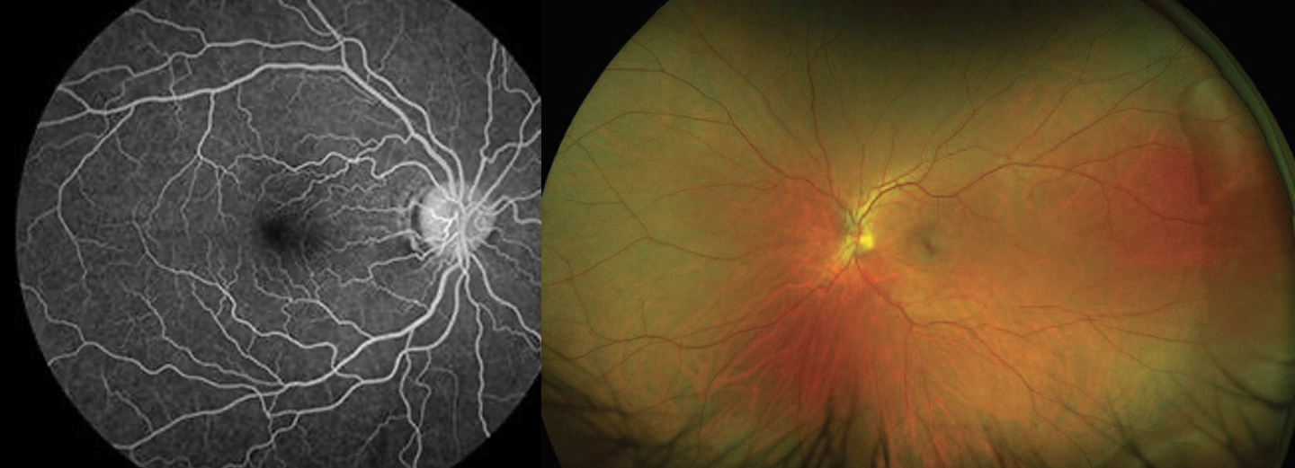
Retinal angiography
Retinal angiography is a minimally invasive eye imaging test that analyzes retinal and choroidal circulation using fundus photographs and dyes. It contributes to the diagnosis of numerous retinal, choroidal and optic nerve pathologies. Your ophthalmologist is willing to answer any additional questions and will provide you with a document summarizing the procedure and its side effects.
What are the stages of the exam?
- Placement of an intravenous line
- Dye injection (fluorescein, indocyanine green, or both)
- Fundus photographs over 5 to 20 minutes
- Ambulatory examination with pupil dilation
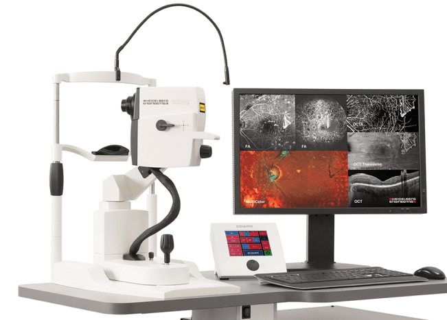
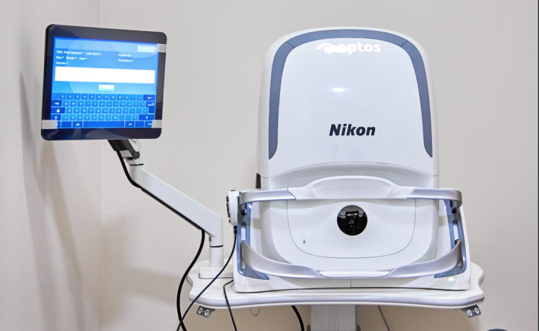
Main indications
- Age-related macular degeneration (AMD)
- Diabetic retinopathy and maculopathy
- Retinal vascular occlusions
- Central serous chorioretinopathy
- Subretinal neovascularization
Autofluorescence
What is autofluorescence?
This non-invasive imaging technique maps the distribution of lipofuscin in pigment epithelial cells, but also other fluorophores that can accumulate pathologically in the retina and subretinal space.
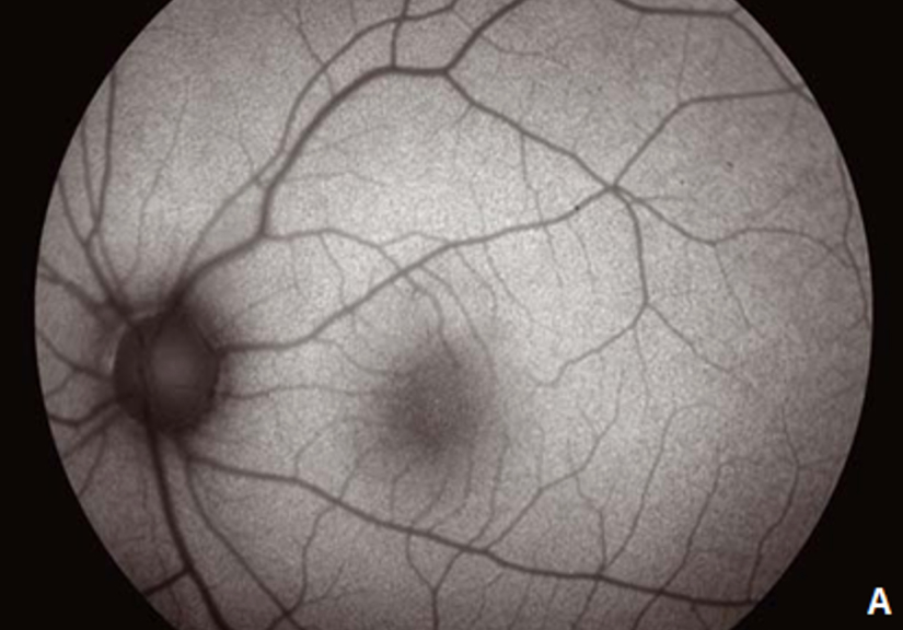
When to perform autofluorescence?
Autofluorescence is useful in cases (non-exhaustive list) of retinopathy, choroidopathy, pseudo-papilloedema and retino-choroidal tumors.
OCT (Optical Coherence Tomography)
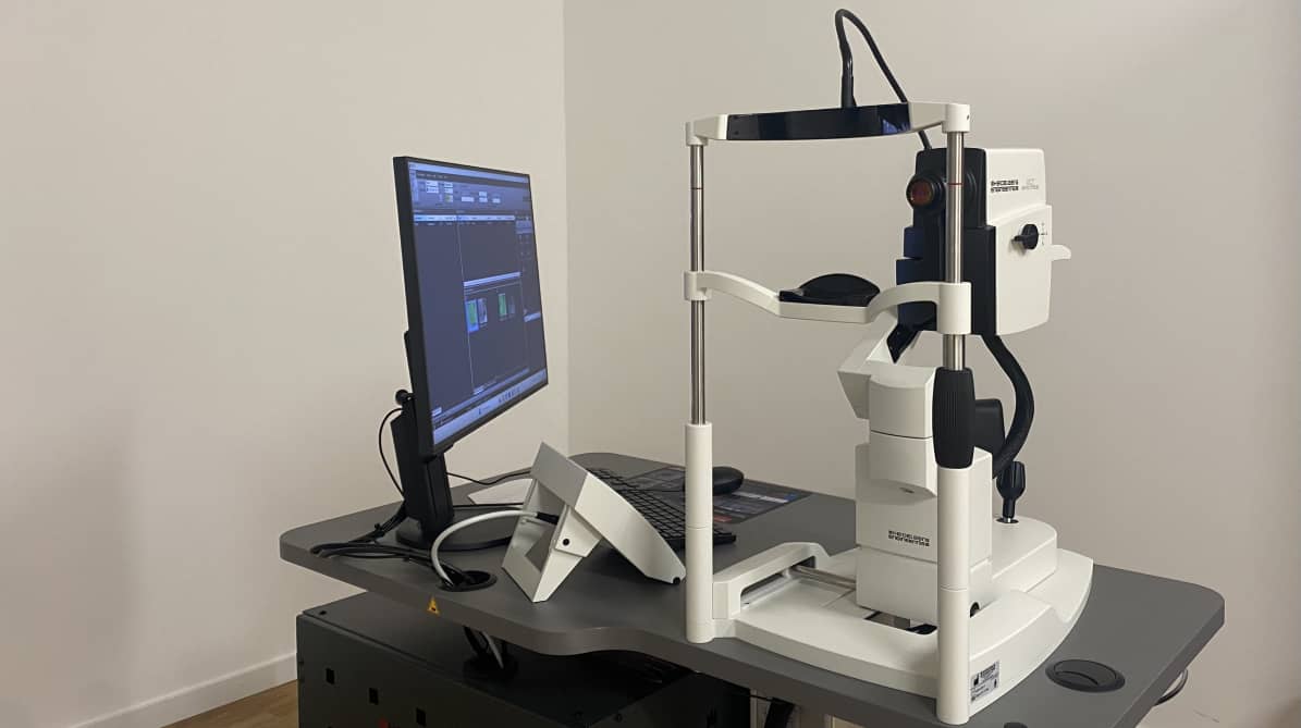
What is OCT?
OCT uses laser interferometry to produce 2D and 3D cross-sectional images without exposure to radiation, providing accuracy comparable to a conventional scanner. It has become an important and precise routine examination in the diagnosis and monitoring of multiple ocular pathologies.
When to perform OCT imaging?
- Macular pathologies (AMD, diabetic maculopathy)
- Peripheral retinal disorders
- Optic nerve assessment (glaucoma)
- Corneal imaging
- Evaluation of the iridocorneal angle