Vitreous & Retina
The retina is the nervous tissue that lines the back of the eye and transforms light into nerve signals. The vitreous is the gel-like substance that fills the back of the eye. Their pathologies can threaten vision and some constitute ophthalmological emergencies.
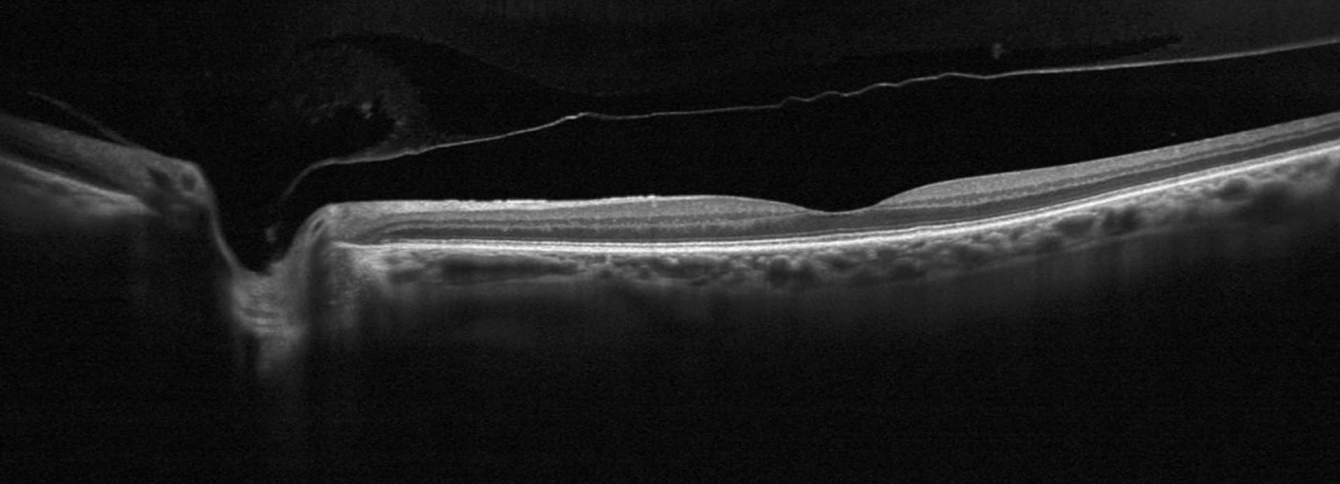
1. Posterior vitreous detachment
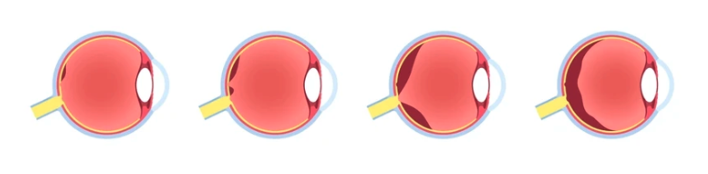
Symptoms of posterior vitreous detachment
What are the common symptoms of PVD?
The vitreous gradually detaches from the retina with age. Typical symptoms are a sudden appearance of floaters (myodesopsia), flashes of light and blurred vision. Although often mild, complications requiring immediate attention can occur.
Treatment of posterior vitreous detachment
What are the therapeutic options?
Uncomplicated PVD does not require treatment. However, monitoring is recommended to detect a possible associated retinal tear.
2. Retinal detachment (rhegmatogenous)
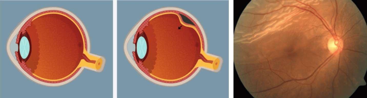
Risk factors for rhegmatogenous retinal detachment
- High myopia
- History of eye surgery
- Eye trauma
- Family history
What are the symptoms of retinal detachment?
Appearance of light flashes, increase in floaters, progressive dark veil in the visual field. Ophthalmological emergency requiring immediate consultation.
What are the treatments for retinal detachment?
- Laser photocoagulation (for tears without separation)
- Pneumatic retinopexy
- Vitrectomy
- Scleral strapping
3. Central serous chorioretinitis (CRSC)
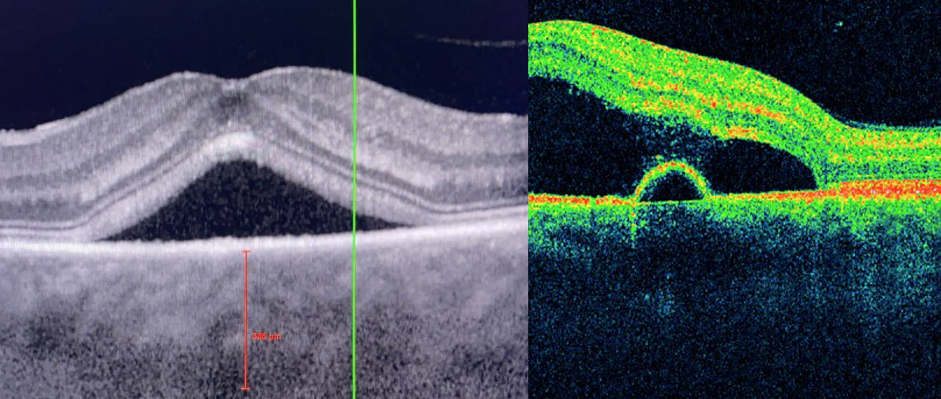
CRSC: what is it?
Accumulation of fluid under the macular retina, often in young men under stress. Causes distorted vision and reduced central acuity.
What are the risk factors?
- Chronic stress
- Corticosteroids (oral, nasal or topical)
- Male, age 20-50 years
What are the symptoms?
Decreased vision, metamorphopsia (distortion of lines), microppsia (objects perceived smaller).
What are the treatments?
Often spontaneous resolution within 3 months. In case of chronic form: thermal laser, PDT (photodynamic therapy) or close observation.
Prevention and Advice
Avoid corticosteroids, reduce stress, maintain a balanced lifestyle.
4. Macular hole
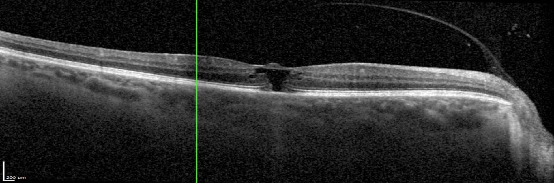
What is a macular hole?
Central perforation of the retina (macula) causing loss of fine central vision. Occurs mainly after age 60.
What are the symptoms?
Dark or blurry spot in the center of the field of vision, distorted straight lines.
What therapeutic options?
Vitrectomy with gas or silicone oil tamponade. High success rate (>90%) with hole closure.
5. Epiretinal membrane
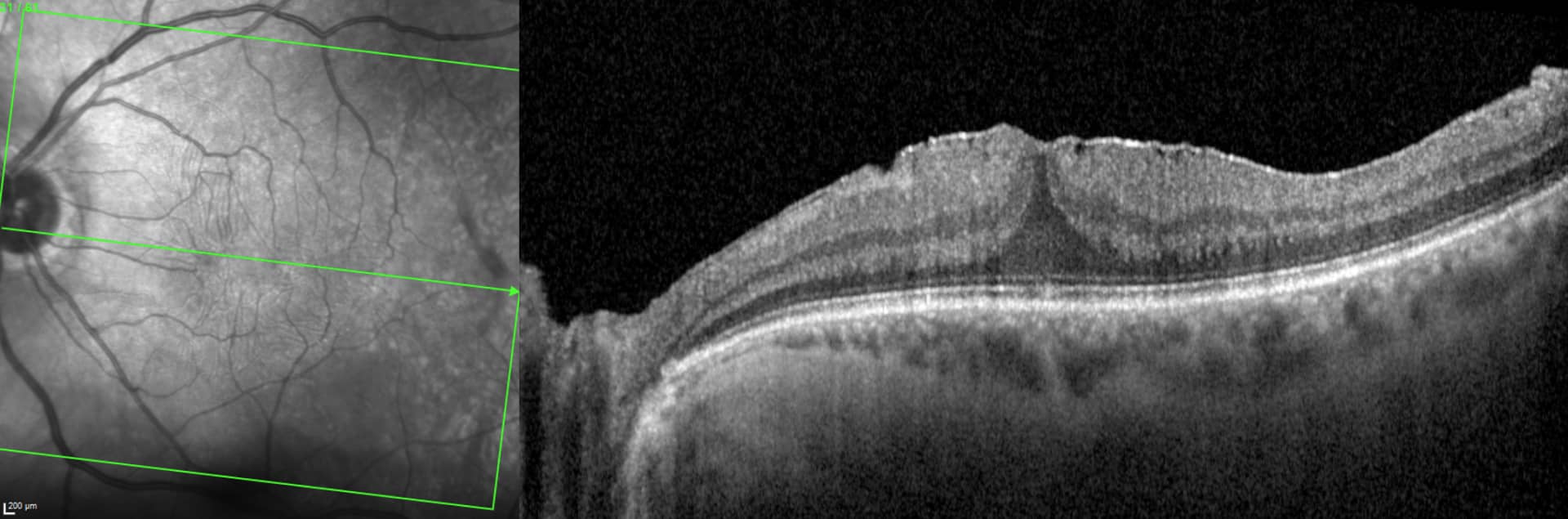
What is an epiretinal membrane?
Fibrous veil that develops on the surface of the macula, causing distortion and distortion of images. May be associated with PVD or inflammation.
What are the treatments?
Monitoring if slight discomfort. In case of significant visual loss: vitrectomy with peeling of the membrane.
6. Retinal vein occlusions (RVO)
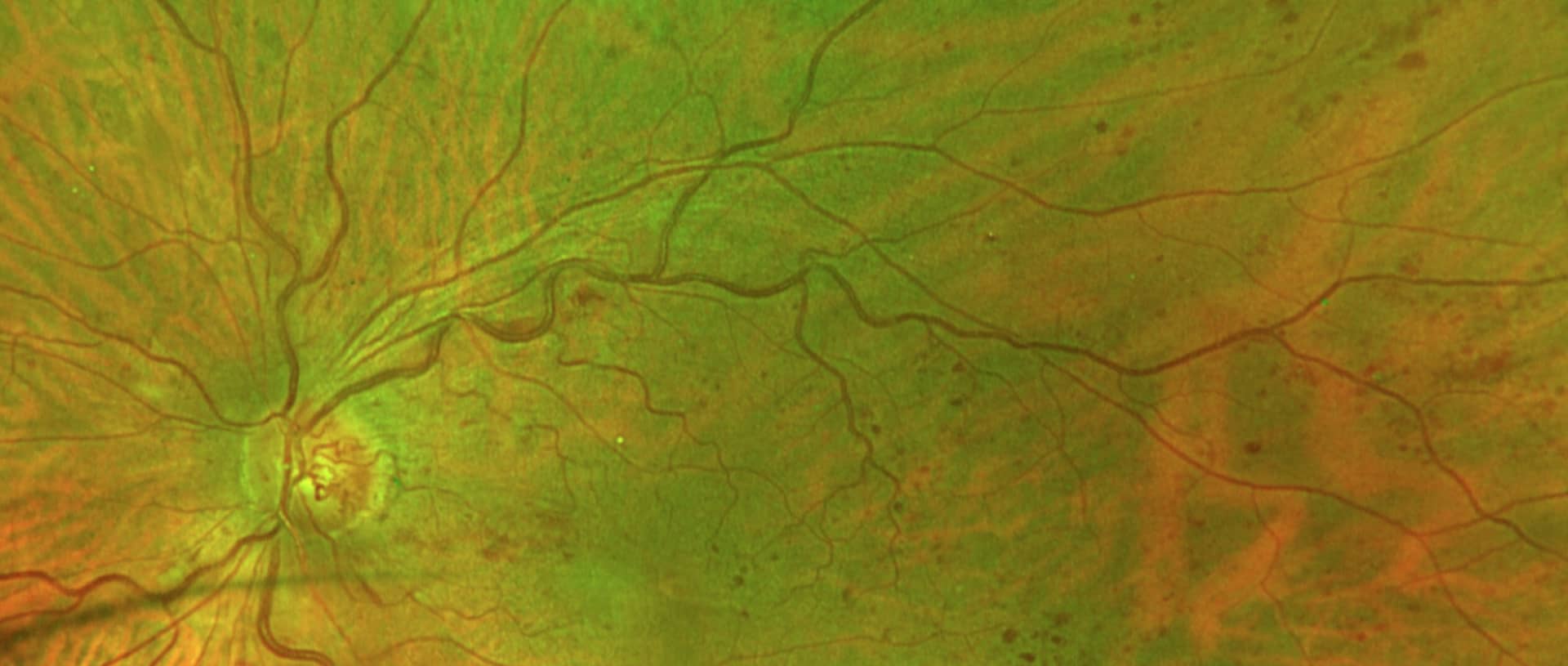
Retinal vein occlusions: what are we talking about?
Obstruction of a retinal vein causing hemorrhages and macular edema in the occluded venous territory. May affect the central vein (CRVO) or a branch (OBVR).
What are the symptoms?
Sudden or progressive loss of vision, visual field deficit.
What are the risk factors?
- High blood pressure
- Diabetes
- Hyperlipidemia
- Glaucoma
7. Retinal Arterial Occlusions (ROA)
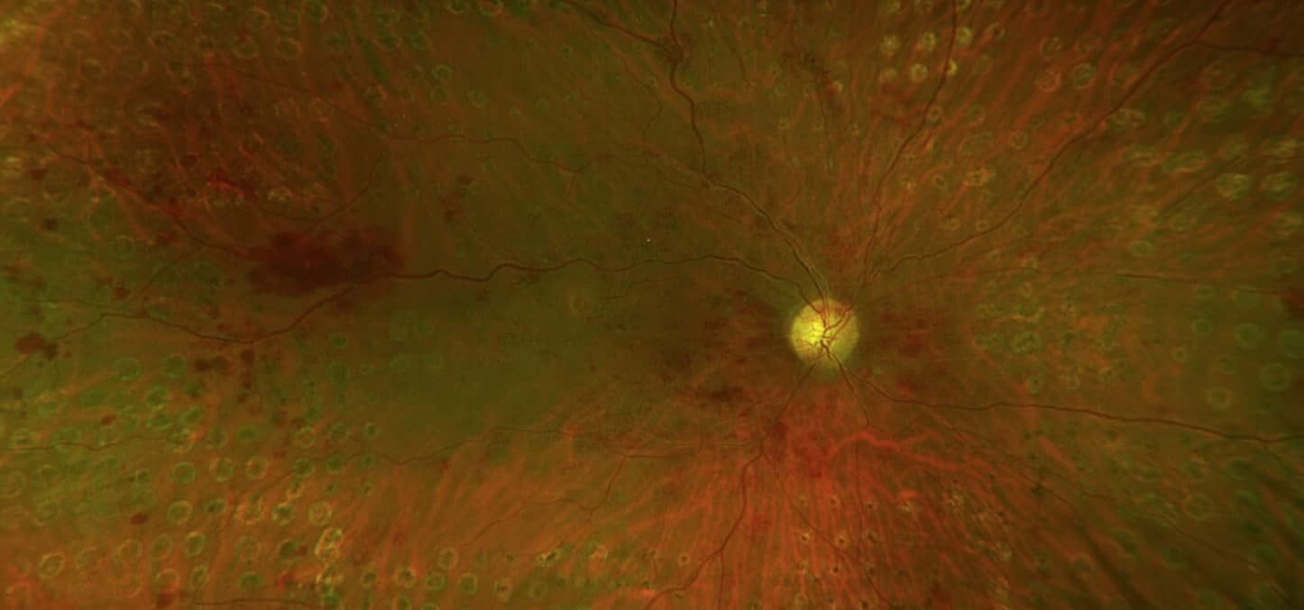
Retinal arterial occlusions: what are we talking about?
Obstruction of a retinal artery leading to immediate ischemia of the retina. Vascular emergency requiring immediate treatment.
What are the symptoms?
Sudden, painless, unilateral or bilateral loss of vision depending on the area affected.
What are the causes and risk factors?
- Embolism (carotid, cardiac)
- Thrombosis
- Giant cell arteritis
- Hypertension, diabetes, bleeding disorders